




Finding out that your baby may have a heart condition can feel overwhelming, particularly if you expected a routine pregnancy scan to provide reassurance. You may suddenly be faced with unfamiliar medical terms, additional appointments and questions about what the findings could mean for your baby’s health.
Although prenatal heart screening has improved significantly, it cannot identify every heart condition before birth. Some abnormalities are difficult to detect during pregnancy, while others become clearer later in pregnancy or only after your baby is born.
What Is Congenital Heart Disease?
Congenital heart disease is a general term for structural problems affecting the heart or major blood vessels that arise while the heart is forming during pregnancy. They may be diagnosed before birth, soon after delivery or later in childhood.
There are many different forms of congenital heart disease, and they can affect the heart in different ways. Some conditions involve a small hole between the heart’s chambers, while others affect the valves, major blood vessels or the development of part of the heart.
The impact on your baby can vary considerably depending on the type and severity of the condition. While some children only need monitoring and regular check-ups, others may require medication, catheter-based treatment or heart surgery to support healthy heart function.
Can Heart Defects Really Be Seen Before Birth?
Yes, many major structural heart defects can be identified during pregnancy. The routine 20-week screening scan assesses specified views of your baby’s heart, while specialist fetal echocardiography can examine its structure, rhythm, function and blood-flow patterns in greater detail.
Early diagnosis can give you time to understand the condition, meet the relevant specialists and plan your baby’s care, although prenatal screening cannot identify every heart condition.
When Does Your Baby’s Heart Develop?
Your baby’s heart begins developing very early in pregnancy. Most of the major structures form during the first trimester, although the heart continues to grow and mature throughout the rest of the pregnancy.
By the time of the 20-week screening scan, sometimes called the anomaly scan, your baby’s heart is usually large enough for the chambers, valves and major blood vessels to be examined in detail.
Some heart abnormalities can be identified relatively early in pregnancy, while others become more noticeable as your baby’s heart continues to develop. This is why you may be offered additional scans or follow-up appointments if further assessment is needed.
The Role of the 20-Week Screening Scan
In England, the NHS Fetal Anomaly Screening Programme offers an ultrasound examination between 18 weeks and 20 weeks plus 6 days of pregnancy, written clinically as 18+0 to 20+6 weeks.
During the scan, the sonographer examines several parts of your baby’s body, including the heart. As part of this assessment, they will look closely at the heart’s structure and screen for a number of specified physical conditions, including some serious congenital heart defects.
The aim of the scan is to identify findings that may require specialist input, additional monitoring during pregnancy or treatment after birth. While the scan provides valuable information about your baby’s development, it is a screening test and cannot detect every possible heart condition.
UK Guidance Note
If the first scan cannot be completed because image quality is compromised, a single repeat scan must be offered and completed by 23+0 weeks.
If image quality is suboptimal and an unexpected structural finding is suspected, a second opinion and referral for further investigation should be arranged without delay rather than waiting for another routine screening appointment.
What Does the Sonographer Look for in the Heart?
The sonographer does much more than simply check whether your baby’s heart is beating. During the scan, a structured assessment is used to examine the position, size, rhythm and overall anatomy of your baby’s heart.
Part of this examination includes a four-chamber view, which allows the two upper chambers and two lower chambers of the heart to be assessed. The sonographer will also examine the major blood vessels leaving the heart and evaluate how structures such as the aorta and pulmonary artery are positioned.
In England, the fetal cardiac protocol includes checking the position of the stomach and heart, known as situs or laterality, as well as the four-chamber view, the ventricular outflow tracts and the three-vessel-and-trachea view.
Screening Is Not the Same as Diagnosis
A routine pregnancy scan is a screening test designed to identify babies who may have an increased likelihood of a health condition. While it can highlight potential concerns, it does not always provide enough information to confirm an exact diagnosis.
In some cases, the sonographer may notice an unusual appearance, an unclear structure or a difference in the size of the heart chambers. If this happens, it does not automatically mean that your baby has a serious heart defect, as there can be several possible explanations for the finding.
You will usually be referred for a specialist assessment before any firm conclusions are made. A fetal cardiologist can examine your baby’s heart in greater detail and explain whether the findings appear normal, remain uncertain or are consistent with a specific heart condition.
What Is a Fetal Echocardiogram?
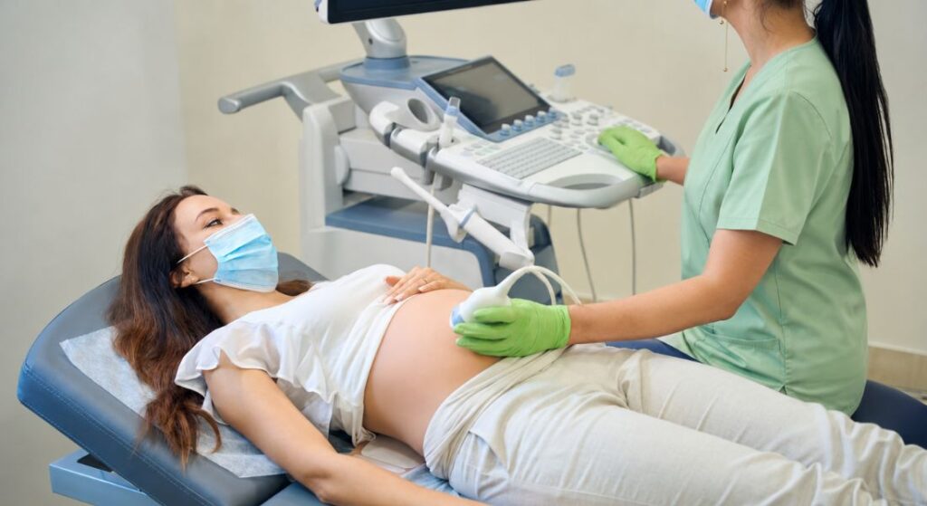
A fetal echocardiogram is a detailed ultrasound scan that focuses specifically on your baby’s heart. It is usually performed by a fetal cardiologist or another specialist with advanced training in fetal heart imaging.
During the examination, the specialist assesses the heart’s chambers, walls, valves, rhythm and major blood vessels. They can also evaluate how blood flows through different parts of your baby’s circulation, helping to build a more complete picture of heart function.
A fetal echocardiogram provides more detailed information than the heart assessment performed during a routine pregnancy scan. It may confirm a suspected heart condition, clarify its severity or reassure you that the original concern is not significant.
| Feature | Routine 20-week screening scan | Specialist fetal echocardiogram |
| Main purpose | Screens the baby’s anatomy, including specified views of the heart | Examines the heart in greater detail when there is a concern or recognised risk factor |
| Typical timing | 18+0 to 20+6 weeks in England | Commonly during the second trimester; earlier or later scans may also be offered |
| Who performs it | An appropriately trained sonographer | A fetal cardiologist or professional with specialist fetal cardiac imaging expertise |
| What is assessed | Cardiac position, four chambers, outflow tracts and major-vessel views | Detailed anatomy, rhythm, function, valves, vessels and blood-flow patterns |
| Result | May appear normal, incomplete or raise a possible concern | May confirm a diagnosis, refine it, recommend follow-up or provide reassurance |
| Limitation | Cannot detect every congenital heart condition | More detailed, but still cannot exclude every small or evolving abnormality |
When Is Fetal Echocardiography Usually Performed?
Detailed fetal cardiac screening is most commonly performed during the second trimester of pregnancy. In many cases, the period between 18 and 22 weeks is considered an ideal time to examine your baby’s heart because the structures are usually large enough to be assessed in detail.
If there is a particular concern, you may be offered a specialist examination earlier in pregnancy. While an early scan can provide valuable information, you may still need a follow-up assessment because your baby’s heart is smaller and some structures can be more difficult to evaluate at that stage.
Additional scans may also be recommended later in pregnancy depending on the findings. Your medical team may arrange further assessments if a condition needs monitoring, if the initial examination was incomplete or if updated information is required before your baby is born.
Why Might You Be Referred for a Specialist Heart Scan?
You may be referred for a specialist heart scan if a routine pregnancy scan identifies a possible abnormality. This could include an unusual four-chamber view, a difference in the size of the ventricles, an abnormal arrangement of the major blood vessels or concerns about your baby’s heart rhythm.
A referral may also be recommended if your pregnancy is associated with a recognised risk factor for congenital heart disease, even when the routine scan appears normal. This allows your baby’s heart to be examined in greater detail and helps ensure that any potential concerns are assessed thoroughly.
Being referred for a fetal echocardiogram does not necessarily mean that a heart defect will be found. In many cases, the specialist examination provides reassurance and confirms that your baby’s heart appears to be developing normally.
How Does Family History Affect the Decision?
A family history of congenital heart disease can increase the likelihood of a baby being born with a heart condition. A specialist fetal heart scan may therefore be considered when either parent or a previous child has congenital heart disease. The importance of the history depends on the exact diagnosis and the referral criteria used by the local fetal cardiology service.
The significance of a family history depends on who has the condition and the specific diagnosis involved. Based on this information, a specialist can assess your baby’s level of risk and decide whether a fetal echocardiogram would be beneficial.
Clinical Tip: Bring Details of the Family Diagnosis
If congenital heart disease affects you, your partner or a previous child, bring any available clinic letters or the exact name of the condition to your appointment. Phrases such as “a hole in the heart” can describe several different defects, so precise information can help the fetal cardiology team assess your baby’s risk and decide which examinations may be appropriate.
Which Maternal Conditions May Lead to Additional Screening?

Certain maternal health conditions may lead to specialist fetal heart screening. These include pre-existing diabetes and, in some autoimmune conditions, the presence of anti-Ro/SSA or anti-La/SSB antibodies. Referral criteria can vary, so your maternity or fetal medicine team will advise whether a detailed fetal heart scan is recommended in your circumstances.
If you have congenital heart disease yourself, your maternity and cardiology teams may advise a fetal echocardiogram. They will also consider how your heart condition could affect your pregnancy, labour and delivery, helping to ensure that both you and your baby receive appropriate care.
Having a recognised risk factor does not mean that your baby will develop a heart defect. It simply means that a more detailed assessment may be helpful, allowing any potential concerns to be identified and managed as early as possible.
Can Medicines or Infections Affect the Fetal Heart?
Only certain medicines and infections are associated with an increased likelihood of fetal heart abnormalities. Whether additional screening is appropriate depends on the specific medicine or infection, the dose, when the exposure occurred and your wider medical history. Your maternity or fetal medicine team can assess these factors individually.
You should not stop any prescribed medication without medical advice. Suddenly discontinuing treatment may affect both your health and your baby’s wellbeing, so it is important to discuss any concerns with the healthcare professional responsible for your care.
Your maternity team may also recommend further assessment if you have experienced a relevant infection during pregnancy. The decision will depend on the type of infection, when it occurred and whether any findings have been seen on your baby’s ultrasound scans.
Which Fetal Findings Can Trigger a Referral?
A referral for a specialist heart scan may be recommended when another structural difference is identified elsewhere in your baby’s body. In some cases, congenital heart conditions can occur alongside abnormalities affecting other organs or as part of a genetic or chromosomal condition.
An increased nuchal translucency measurement in early pregnancy may also lead to a detailed cardiac assessment, even if later screening results appear reassuring. Other reasons may include an abnormal fetal heart rhythm, fetal hydrops or significant fetal growth restriction, particularly when there are additional ultrasound findings or recognised risk factors. Whether a specialist heart scan is recommended will depend on the complete clinical picture and local referral criteria.
These findings do not necessarily mean that your baby has a heart condition. Instead, they suggest that a more focused examination of the heart may provide valuable information and help clarify whether any further monitoring or treatment is needed.
What Happens During the Fetal Heart Scan?
A fetal heart scan is performed in a similar way to other ultrasound examinations during pregnancy. You will usually lie on an examination couch while gel is applied to your abdomen, allowing the specialist to move the ultrasound probe across your skin and obtain detailed images of your baby’s heart.
During the scan, the specialist examines the heart from several different angles to assess its structure and function. You may be asked to change position, take a short walk or wait for a while if your baby needs to move into a better position for imaging.
The appointment often takes longer than a routine pregnancy scan because the heart is examined in much greater detail. The length of the scan can vary depending on your baby’s position, the quality of the images obtained and whether any findings require closer assessment.
How Is Blood Flow Examined?
The specialist may use Doppler ultrasound to assess how blood moves through your baby’s heart, valves and major blood vessels. Colour Doppler can show the direction of blood flow, while other Doppler techniques help measure its speed and pattern.
This information helps the fetal cardiologist understand whether the heart valves are functioning properly and whether blood is flowing through the expected pathways. It can also help identify problems such as narrowing, leakage or unusual connections within the heart or blood vessels.
Although the images and terminology may appear highly technical, the specialist should explain the key findings in a clear and understandable way. You should feel comfortable asking questions if there is anything you do not understand or if a medical term needs further explanation.
Is Fetal Echocardiography Safe?
Fetal echocardiography is a non-invasive examination performed through your abdomen. It uses ultrasound rather than X-rays or other forms of ionising radiation.
No confirmed harmful effects have been demonstrated when diagnostic ultrasound is used appropriately by trained professionals. Specialists use the lowest ultrasound output and shortest examination time needed to obtain the required information. In most cases, you can eat, drink and take your usual medication before the appointment unless your clinic advises otherwise.
Why Are Some Defects Missed Before Birth?
Some heart defects are difficult to identify before birth because the fetal circulation works differently from the circulation after delivery. Certain natural connections remain open while your baby is in the womb, which can make some abnormalities harder to recognise during pregnancy.
Very small openings between the lower chambers of the heart may be too small to be seen on prenatal ultrasound. In addition, some conditions involving the pulmonary veins, aortic arch or smaller heart valves can be challenging to diagnose reliably before birth.
The quality of the images can also be affected by factors such as your baby’s position, movement, gestational age or other technical considerations. As a result, a heart may appear normal during pregnancy, but a condition can sometimes become apparent only after your baby is born.
Why Might You Need a Repeat Scan?
A repeat scan may be recommended if the specialist was unable to obtain all the views needed to assess your baby’s heart properly. This can happen when your baby is in an awkward position or moving frequently during the examination, making certain structures difficult to see clearly.
Being asked to return for another scan does not necessarily mean that there is a problem. In many cases, a follow-up appointment is arranged simply because the required screening views could not be completed during the initial examination.
Additional assessment becomes particularly important if an unexpected finding is suspected. If there is a concern about your baby’s heart, you may be referred for specialist evaluation without waiting for another routine screening appointment, allowing any potential issue to be investigated more promptly.
Which Heart Conditions Can Be Detected?
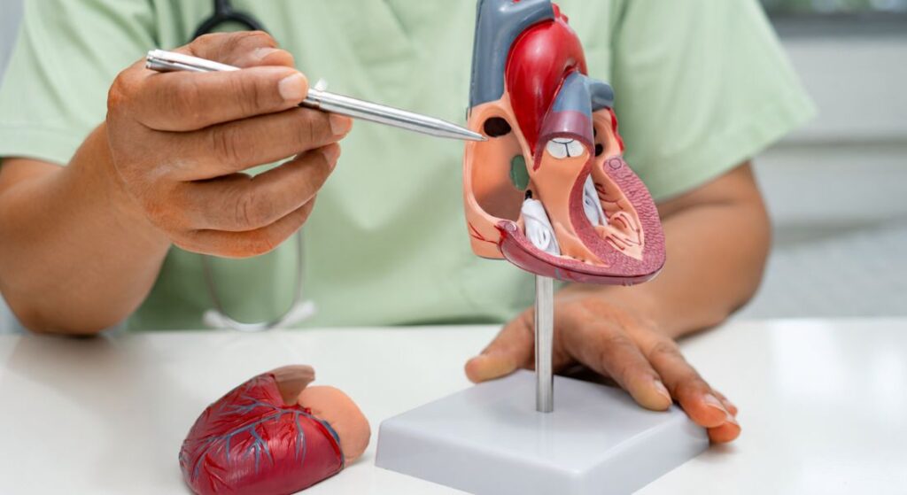
Prenatal ultrasound can identify many major structural heart abnormalities before birth. These may include conditions such as hypoplastic left heart syndrome, transposition of the great arteries, tetralogy of Fallot and significant problems affecting the heart valves or major blood vessels.
The scan may also detect some ventricular septal defects, which are openings between the lower chambers of the heart, abnormalities involving the atrioventricular valves and conditions where only one main pumping chamber has developed. However, some heart defects are easier to identify than others, and certain conditions may become more apparent as your baby’s heart continues to develop.
Prenatal detection is particularly important when a baby may need specialist care soon after birth. It allows maternity, neonatal and cardiac teams to plan the place of delivery and arrange timely assessment or treatment.
Evidence Note
Research, including a meta-analysis of observational studies, suggests that prenatal diagnosis can improve delivery and neonatal-care planning for babies with critical congenital heart disease.
Among selected babies with comparable cardiac anatomy whose families planned surgical treatment, prenatal diagnosis was associated with a lower risk of death from cardiovascular compromise before planned neonatal surgery. However, this evidence does not establish the same benefit for every heart defect and cannot predict an individual baby’s outcome.
The main established benefit of prenatal detection is that maternity, neonatal and cardiac teams have time to plan the place of delivery, immediate assessment and any treatment that may be required after birth.
Can a Heart Condition Change During Pregnancy?
Some fetal heart conditions remain relatively stable throughout pregnancy, while others can change as your baby continues to grow and develop. In some cases, narrowing of a valve, differences between the ventricles or obstruction within a major blood vessel may become more noticeable on later scans.
For this reason, your fetal cardiologist may recommend regular follow-up appointments. These scans allow the team to monitor your baby’s heart development, assess its rhythm and evaluate how well it is functioning over time.
Ongoing assessments also help specialists refine the diagnosis and plan the most appropriate care before and after birth. As new information becomes available, you may find that the expected outlook or delivery plan is updated to reflect the latest findings.
Will Genetic Testing Be Recommended?
Some congenital heart conditions occur on their own, while others are linked to chromosomal or genetic differences. The likelihood depends on the specific heart defect identified and whether any additional abnormalities have been seen on your baby’s ultrasound scans.
If further assessment is recommended, you may be offered an appointment with a fetal medicine specialist or genetic counsellor. Non-invasive prenatal testing, commonly called NIPT, is a screening test for selected chromosomal conditions and cannot diagnose every genetic cause of congenital heart disease.
Chorionic villus sampling and amniocentesis are diagnostic procedures that can test for chromosomal or genetic conditions selected by your specialist team. However, even diagnostic testing cannot identify every possible genetic cause of congenital heart disease.
The decision about genetic testing is always yours to make. Your healthcare team should explain what each test can detect, its limitations, how long the results may take and whether the findings could influence your pregnancy management or your baby’s care after birth.
Who Will Be Involved in Your Care?
Your care may involve several healthcare professionals working together to support both you and your baby throughout the pregnancy. Depending on the diagnosis, this team may include a fetal cardiologist, fetal medicine specialist, obstetrician, midwife, genetic specialist, neonatal doctor and members of the children’s cardiac team.
The professionals involved will vary according to the type of heart condition and the level of care your baby may need after birth. If early treatment is likely to be required, coordinated care becomes particularly important to help ensure that appropriate plans are in place before delivery.
Communication between specialists is an important part of this process. Reports from your baby’s heart scans are usually shared with your maternity team so that everyone involved in your care has the same information and can provide consistent advice.
How Will the Severity of the Condition Be Explained?
The name of a heart condition does not always indicate exactly how it will affect your baby. To provide a clearer picture, the specialist will assess the heart’s anatomy, blood flow, overall function and whether any other medical findings are present.
You should be given information about the likely severity of the condition and what it may mean after birth. This may include whether your baby is expected to need monitoring, treatment later in childhood or specialist care soon after delivery, while also discussing any uncertainties that remain.
It can be helpful to ask the specialist to explain the findings using a simple diagram. You may also wish to request a written summary, as it can be difficult to remember detailed information when you are processing a significant amount of new information.
What Happens Immediately After a Prenatal Diagnosis?
Following a prenatal diagnosis, the specialist will explain what has been identified and how confident they are in the findings. You will usually have an opportunity to ask questions and take time to understand the information before discussing the likely outlook and next steps.
If the condition is serious, the fetal medicine team should provide balanced and non-directive information about all options available to you. Depending on the diagnosis and stage of pregnancy, this may include continuing the pregnancy with a detailed care plan or discussing the option of ending the pregnancy. You should be given time to ask questions, involve people you trust and access emotional or counselling support.
Further scans may be arranged to monitor your baby’s heart and assess their ongoing development throughout the pregnancy. Depending on the findings, you may also be offered genetic assessment or the chance to meet members of the neonatal and children’s cardiac teams before delivery.
A prenatal diagnosis provides valuable time for planning and preparation. In most cases, congenital heart conditions are managed after birth, so a diagnosis during pregnancy does not necessarily mean that your baby will need treatment before delivery.
Will the Diagnosis Affect Where Your Baby Is Born?
Some babies with less complex congenital heart disease may be born at their local maternity unit and assessed by specialists after delivery. However, the recommended place of birth depends on the diagnosis, the expected condition of the baby after birth and how quickly specialist treatment may be required.
If urgent treatment or specialist care is expected, you may be advised to give birth at or near a specialist cardiac centre. This can help ensure that your baby has rapid access to medication, neonatal support, intensive care or a children’s cardiac team if needed.
Your maternity and cardiac specialists will work together to create an individual birth plan for you and your baby. This plan should outline the recommended hospital, the support available after delivery and what arrangements will be made if labour starts earlier than expected.
Will You Need a Caesarean Section?
A prenatal diagnosis of congenital heart disease does not automatically mean that you will need a caesarean section. Vaginal birth remains suitable in many cases unless there is a separate obstetric reason or a condition-specific clinical reason to recommend a different approach.
Your obstetric and fetal cardiology teams will consider your health, your baby’s condition, the progress of the pregnancy and access to specialist care. They should explain the reasons, benefits and risks if induction or caesarean birth is recommended.
What About Future Pregnancies?

If your baby is diagnosed with congenital heart disease, it is natural to wonder whether the condition could affect future pregnancies. The likelihood of recurrence depends on the specific diagnosis, your family history and whether a genetic cause has been identified.
Your healthcare team may recommend genetic counselling before or during another pregnancy. This can help you understand any potential risks and provide more personalised guidance based on your family’s medical history and previous test results.
In a future pregnancy, you may be offered earlier screening and specialist fetal echocardiography to assess your baby’s heart in greater detail. Having a clear plan in place can provide reassurance and help ensure that any necessary investigations are arranged at the most appropriate stage of pregnancy.
What Treatment Might Your Baby Need After Birth?
After birth, your baby may have an echocardiogram to confirm the findings seen during pregnancy. Doctors may also check oxygen levels, perform an electrocardiogram (ECG) and monitor your baby’s breathing, feeding and circulation during the first days of life.
The treatment required will depend on the specific heart condition and its severity. Some babies need only observation and regular follow-up appointments, while others may require medication to support circulation or keep an important blood vessel called the ductus arteriosus open until further treatment is possible.
In some cases, treatment may involve a catheter-based procedure or heart surgery. The timing can vary considerably, with some babies needing intervention within the first few hours or days after birth, while others may not require treatment until months or even years later.
What Does a Normal Fetal Echocardiogram Mean?
A normal fetal echocardiogram is reassuring because it makes a major structural heart abnormality much less likely. It means that your baby’s heart structure, rhythm, function and blood-flow patterns appeared appropriate at the time of the examination.
However, a normal result cannot exclude every possible heart condition. Some small septal defects, mild valve abnormalities and conditions that develop or become more apparent later may not be detectable before birth.
Call 999 if your baby’s lips, tongue, face or skin suddenly turn blue or grey, or if your baby has severe difficulty breathing, becomes floppy or is difficult to wake. On brown or black skin, a change in colour may be easier to see on the lips, tongue, gums, palms, soles or inside the eyelids. Ask for an urgent GP appointment or contact NHS 111 if your baby has persistent feeding difficulties, unusual breathlessness or poor weight gain.
Even when prenatal scans are normal, your baby should still have the routine newborn physical examination after birth. This remains important because some congenital heart conditions are small, develop later or become apparent only when the circulation changes after delivery.
Myth vs Fact
| Myth | Fact |
| A normal 20-week scan rules out every heart defect. | A normal result is reassuring, but small, evolving or difficult-to-image abnormalities may not be detected before birth. |
| Referral for fetal echocardiography means a heart defect has definitely been found. | Many referrals are precautionary, and the specialist scan may confirm that the heart appears normal. |
| Fetal echocardiography uses radiation. | It uses ultrasound rather than ionising radiation. |
| A prenatal CHD diagnosis always means the baby will need immediate surgery. | Some babies need monitoring only, while others may need medication, a catheter procedure or surgery at different stages. |
| A prenatal cardiac diagnosis automatically requires a caesarean section. | Vaginal birth is suitable in many cases unless there is an obstetric or condition-specific reason for a different delivery plan. |
| Every congenital heart defect can be detected during pregnancy. | Detection depends on the type of defect, gestational age, image quality, fetal position and whether the condition develops or becomes clearer later. |
Key Takeaways
- Many significant congenital heart defects can be identified during pregnancy, but prenatal screening cannot detect every condition.
- In England, the NHS 20-week screening scan is offered between 18+0 and 20+6 weeks.
- Fetal echocardiography provides a more detailed assessment of the heart’s anatomy, rhythm, function and blood flow.
- Referral for specialist assessment does not necessarily mean that a heart defect has been confirmed.
- Prenatal diagnosis can help specialists plan delivery, neonatal assessment and treatment where required.
- A normal fetal echocardiogram makes a major structural abnormality less likely but cannot exclude every small or evolving defect.
- Genetic testing may be offered depending on the cardiac diagnosis and any additional ultrasound findings.
- After birth, call 999 if your baby’s lips, tongue, face or skin suddenly turn blue or grey, or if your baby has severe difficulty breathing, becomes floppy or is difficult to wake.
Frequently Asked Questions
1. Can congenital heart disease be detected before birth?
Yes, many forms of congenital heart disease can be identified during pregnancy through routine ultrasound screening and specialist fetal heart scans. The 20-week screening scan can detect a number of significant heart abnormalities, although some conditions may require further assessment with fetal echocardiography. Early diagnosis allows healthcare professionals to plan care before and after birth. However, not all heart defects can be detected during pregnancy.
2. What is a fetal echocardiogram?
A fetal echocardiogram is a detailed ultrasound examination that focuses specifically on your baby’s heart. It is usually performed by a fetal cardiologist or another specialist trained in fetal heart imaging. The scan assesses the heart’s structure, valves, chambers, blood vessels and blood flow patterns. It provides more detailed information than a routine pregnancy ultrasound.
3. Why might a specialist fetal heart scan be recommended?
You may be referred for a fetal echocardiogram if a routine pregnancy scan raises concerns about your baby’s heart. Other reasons include a family history of congenital heart disease, certain maternal medical conditions or findings elsewhere on your baby’s ultrasound scans. In many cases, the specialist scan provides reassurance that the heart is developing normally. A referral does not automatically mean that a heart defect has been found.
4. At what stage of pregnancy can congenital heart disease be detected?
Many significant heart defects can be identified during the second trimester, particularly between 18 and 22 weeks of pregnancy. Some abnormalities may be visible earlier, while others become easier to detect as the heart continues to develop. Depending on the findings, additional scans may be recommended later in pregnancy. The timing of detection often depends on the type of condition and the quality of the images obtained.
5. Is fetal echocardiography safe for me and my baby?
Fetal echocardiography is a non-invasive examination that uses ultrasound rather than ionising radiation. No confirmed harmful effects have been demonstrated when diagnostic ultrasound is used appropriately by trained professionals. Your clinic will tell you if any preparation is required.
6. Can a normal pregnancy scan guarantee that my baby does not have a heart condition?
No, a normal pregnancy scan is reassuring but cannot completely rule out every heart condition. Some defects are very small or difficult to identify before birth because the fetal circulation works differently from circulation after delivery. Certain abnormalities may only become apparent later in pregnancy or after your baby is born. This is why routine newborn examinations remain important even when prenatal scans appear normal.
7. What happens if congenital heart disease is diagnosed during pregnancy?
If a heart condition is identified, your specialist team will explain the diagnosis, discuss its severity and outline the likely next steps. You may be offered additional scans, genetic counselling or meetings with specialists who will be involved in your baby’s care. A personalised plan can then be developed for the remainder of the pregnancy, delivery and postnatal care. Having this information before birth can help families prepare and ensure appropriate support is available.
8. Will a prenatal diagnosis affect where I give birth?
The answer depends on the type and severity of the heart condition. Many babies with congenital heart disease can be delivered at their local maternity unit and reviewed by specialists after birth. If urgent treatment is expected, your healthcare team may recommend giving birth at or near a specialist cardiac centre. This helps ensure your baby has immediate access to the care they may need.
9. Does a prenatal diagnosis mean my baby will need surgery after birth?
Not necessarily. Some babies with congenital heart disease only require monitoring and regular follow-up appointments, while others may need medication, catheter-based procedures or surgery. The treatment plan depends on the specific heart condition and how it affects your baby’s circulation and heart function. Your specialist team will explain the likely treatment options based on the diagnosis.
10. Can congenital heart disease affect future pregnancies?
Having had a pregnancy affected by congenital heart disease can increase the likelihood of congenital heart disease in a future pregnancy. However, the degree of risk varies considerably according to the specific diagnosis, family history and whether an underlying genetic cause has been identified.
Final Thoughts: The Importance of Early Detection in Congenital Heart Disease
Advances in prenatal screening and fetal echocardiography mean that many heart conditions can now be identified before a baby is born. Although not every cardiac abnormality can be detected during pregnancy, an early diagnosis can provide valuable time to understand the condition, meet the appropriate specialists and plan for your baby’s care after delivery. This preparation can support coordinated delivery planning, timely specialist assessment and early treatment when it is needed.
It is also important to remember that a prenatal diagnosis does not automatically mean your baby will require immediate surgery or experience severe health problems. Many children with congenital heart disease can live active and fulfilling lives, although their health needs and long-term outlook depend on the type and severity of their condition. Ongoing advances in fetal medicine, paediatric cardiology and cardiac surgery continue to improve outcomes for children born with heart defects.
If a possible heart abnormality is identified during pregnancy, your maternity or fetal medicine team should arrange referral to an appropriate fetal cardiology service. After your baby is born, specialist paediatric assessment and ongoing follow-up may be needed depending on the diagnosis.
If your child needs specialist paediatric care for congenital heart disease after birth, you can contact London Paediatric Clinic for assessment, guidance and ongoing support.
References:
- British Congenital Cardiac Association (2021) Fetal Cardiology Standards. Revised September 2021. Available at: https://www.bcca-uk.org/fetal-cardiology
- Carvalho, J.S. et al. (2023) ‘ISUOG Practice Guidelines (updated): fetal cardiac screening’, Ultrasound in Obstetrics & Gynecology, 61(6), pp. 788–803. Available at: https://pubmed.ncbi.nlm.nih.gov/37267096/
- Holland, B.J., Myers, J.A. and Woods, C.R. Jr (2015) ‘Prenatal diagnosis of critical congenital heart disease reduces risk of death from cardiovascular compromise prior to planned neonatal cardiac surgery: a meta-analysis’, Ultrasound in Obstetrics & Gynecology, 45(6), pp. 631–638. Available at: https://pubmed.ncbi.nlm.nih.gov/25904437/
- Moon-Grady, A.J. et al. (2023) ‘Guidelines and recommendations for performance of the fetal echocardiogram: an update from the American Society of Echocardiography’, Journal of the American Society of Echocardiography, 36(7), pp. 679–723. Available at: https://pubmed.ncbi.nlm.nih.gov/37227365/
- NHS (2023) ‘Blue or grey skin or lips (cyanosis)’. Page last reviewed 4 October 2023. Available at: https://www.nhs.uk/symptoms/blue-skin-or-lips-cyanosis/
- NHS (2024) ‘20-week screening scan’. Page last reviewed 1 October 2024. Available at: https://www.nhs.uk/pregnancy/your-pregnancy-care/20-week-scan/
- NHS (2025) ‘Congenital heart disease’. Page last reviewed 11 December 2025. Available at: https://www.nhs.uk/conditions/congenital-heart-disease/
- NHS England (2026a) ‘20-week screening scan’, NHS Fetal Anomaly Screening Programme handbook. Updated 15 July 2026. Available at: https://www.gov.uk/government/publications/fetal-anomaly-screening-programme-handbook/20-week-screening-scan
- NHS England (2026b) ‘Prenatal diagnosis’, NHS Fetal Anomaly Screening Programme handbook. Updated 15 July 2026. Available at: https://www.gov.uk/government/publications/fetal-anomaly-screening-programme-handbook/prenatal-diagnosis
- Sun, H.Y. (2021) ‘Prenatal diagnosis of congenital heart defects: echocardiography’, Translational Pediatrics, 10(8), pp. 2210–2224. Available at: https://pmc.ncbi.nlm.nih.gov/articles/PMC8429868/